Room Temperature Semiconductor Detector Conference
05 – 12 November 2022, Milano, Italy
The IEEE Nuclear Science Symposium (NSS) 2022 will bring together the very large international community of detector scientists and engineers in Milano, Italy. The NSS 2022 program incorporates the latest developments in detector technology and materials, new instrumentation techniques, their implementation in high energy and nuclear physics, astrophysics, accelerators, national nuclear security, and many other applications in various types of radiation environments and emerging fields like quantum science, as well as state-of-the-art developments included in the joint sessions with the MIC and RTSD. Special topic workshops will cover areas of specific interests. Short courses on a variety of traditional as well as novel topics of interest proposed by the NSS community will also be offered.
Authors are invited to submit papers describing their original, unpublished work on one of the topics below:
Brian P. Gaucher For the last six years Brian Gaucher has been a Principle RSM (Research Staff Member) / manager and sub-theme technical lead of IBM’s quantum computing future systems design at IBM research, where his work is focused on novel architectures for quantum electronics for scientific and commercial applications. Specifically, from 2019-2022 he managed two Quantum gates teams and co-lead of the custom cryo-CMOS control and readout development. From 2016-2019 he led the development and implementation of IBM’s room temperature based control electronics for Quantum systems and deployment. Previously he was Sr. Manager /Principal RSM of IBM’s Cognitive Environments Lab focused on the creation and the application of AI in highly interactive physical spaces designed to improve decision-making through always-on ambient intelligence; a human machine partnership to enhance cognition. He’s been an IBM master inventor with over 60 US and foreign patents pending, filed or issued. He’s received two Corporate Awards, three Outstanding Technical Achievement awards, an Outstanding Innovation award, a Research Division Award and has authored or co-authored over 50 papers and one book.
Quantum computers hold the promise to address some of the world’s most important, complex and challenging problems of our times. Currently, the state of the art of Quantum computing is a careful melding of cryogenic quantum processors with classical electronics in the hundreds of qubits scale. Within a few short years, we will likely see advances that push us towards 10’ks to 100’ks of physical qubit sized quantum processors, which will open the door to the possibility of fault tolerant computing, and the ability to address a segment of those most complex problems. This progress in quantum processors is driving the need to create highly scalable, more integrateable and lower cost electronics to operate these systems. Teams around the world are now beginning to leverage CMOS to mitigate these issues and maintain the required performance and to move the electronics into the dilution fridge. That suggests the need for cryoCMOS to address the scaling, integration and costs issues, all while maintaining similar performance, yet at orders of magnitude lower cost, smaller size, lower power consumption and power dissipation, which will drive major innovation in low power CMOS circuit design and implementation at cryogenic temperatures. This talk will provide a simple overview of what Quantum computing is, it will motivate the discussion with a few of the major challenges, discuss what the current state of the art is and where we are in that journey to cryoCMOS Quantum computing control and readout.
Jean-Francois Pratte received his M.A. Sc. in electrical engineering at Université de Sherbrooke, Sherbrooke, Canada in 2002. He then held a position as a research engineer at the Instrumentation Division of the Brookhaven National Laboratory (BNL, U.S. DOE) and obtained in parallel his Ph.D. in 2008 for the realization of the RatCAP (Rat Conscious Animal PET scanner). He was a key designer in the implementation of the first dual modality PET and MRI scanner. While at BNL, he coauthored 3 patents—all transferred to the industry.
In 2009, he joined the Electrical and Computer Engineering Department at Université de Sherbrooke where he currently holds a professor position. His research interests target microsystem design and integration of Photon-to-Digital Converters, where an array of single photon avalanche diodes is integrated vertically in 3D with a pixelated CMOS readout circuit. His current research areas are radiation instrumentation for neutrino physics, dark matter search, medical imaging, as well as quantum key distribution receivers and time-to-digital converters. He was awarded the 2018 Radiation Instrumentation Early Career Award of the IEEE Nuclear and Plasma Science Society “For Spearheading the Development of per Pixel Picosecond Timing with Single Photon Avalanche Diodes Three-Dimensionally Integrated to Custom Readout Circuits”.
Analog SiPMs are used in many scientific experiments and medical imaging systems and are planned as photon sensors for experiments currently under design or construction. Digital SiPM share the same basic photon sensor: a Single Photon Avalanche Diode (SPAD), but in these devices, the “digital” attribute addresses important limitations of their analog counterparts. Thanks to progress in the semiconductor industry, digital SiPM can now be vertically integrated over a pixelated readout electronics. This led to front and backside illuminated 3D digital SiPM, a.k.a. Photon-to-Digital Converters (3D PDC). These, thanks to their enhanced performance, can fulfill an extended set of requirements as it will be discussed.
Several experiments have shown interest in the advanced performance of 3D PDC devices; thus, this presentation is timely in updating scientists on envisioning what may be possible.
A review of the latest advancements in the academic and industrial side will be discussed to help understand where the field is heading and foresee what can be achieved with these new devices.
Digital SPAD arrays are also used to measure charged particles. In some cases, they will be exposed to significant radiation doses from various particles. This aspect will be presented as well.
The final objective of this talk is to get more traction in our scientific community to stimulate the development of these technologies. This requires scientists at all stages and from various fields of expertise, from theoretical physicists who specify the required performance, to experimentalists who are willing to “play” with these devices, to engineers designing systems. But most importantly, this field requires brilliant early career/graduate students to bring new ideas and skills for the benefit of our community and the scientific discoveries they can enable.
Alessandro (“Sandro”) Olivo is a Professor of Applied Physics with the Department of Medical Physics and Biomedical Engineering at University College London. He has worked in x-ray imaging and detection for >25 years, and is considered a pioneer in the field of phase-based x-ray imaging, having co-designed the in vivo mammography system operational at the Trieste synchrotron and invented the “edge-illumination” and “coded-aperture” methods. He founded the UCL Advanced X-Ray Imaging (AXIm) Group in 2008; AXIm has since become a multi-PI consortium with 30 members and in excess of 50 external collaborators across academia, research institutes and industry. In 2019 he was awarded a Chair in Emerging Technologies by the Royal Academy of Engineering, which releases him from teaching and admin duties and allows him to concentrate on technological development.
The use of x-ray imaging is ubiquitous in science and society – most people associate it with medical imaging and possibly security inspections, but x-rays are widely used in many other fields encompassing biology, metrology, industrial inspections, cultural heritage preservation and others. Innovations developed over the last couple of decades – notably phase-based x-ray imaging, detection of x-rays scattered in the microradian range and “colour” (energy-resolved) x-ray imaging are slowly finding their way into mainstream applications. My group’s main contribution to this area of research was the development of methods that enable the implementation of phase-based x-ray imaging and the detection of microradian scattering with conventional, rotating anode x-ray sources, without the need for micro-focal sources or forms of source collimation that inevitably limit the available flux, leading to exposure time increases. Following early proof-of-concept studies these methods are now being translated into medical, industrial and security applications. This talk will briefly discuss the methods’ basic principles, describe their evolution from laboratory proof-of-concept to pre-commercial prototype, present a series of the most recent applications in the above fields, and discuss interactions with other quickly developing areas such as colour x-ray imaging and machine learning.
The IEEE Medical Imaging Conference (MIC) is a leading international scientific meeting to discuss the latest physics, engineering, and mathematical aspects of medical imaging with a particular focus on ionization radiation.
Medical imaging is a continuously growing field while detectors, instrumentation, modern computational methods, and integrated systems are leading the way towards technological advances
MIC has a unique focus on cutting edge technologies as well as their effective translation to clinical practice. In recent years, interest has increased in applications of machine learning, AI, and other rapidly emerging areas of research which will be also dealt with. Physics and engineering solutions for medical unmet needs are another important topic.
MIC is an opportunity for students, post-doctoral fellows, and junior and senior researchers from around the world to come together and share their new ideas and results of innovations and scientific endeavors.
The scientific program of the MIC consists of oral and poster sessions, plenary sessions, and a student award session. Regular sessions will be complemented by Short Courses and specialized Workshops covering timely topics in medical imaging and therapy.
Cyril Lafon earned his B.Sc. degree in Physics from University Blaise Pascal of Clermont Ferrand, France and University of Montreal, Quebec, in 1995, and his Ph.D. degree in Biomedical Engineering from University Claude Bernard of Lyon, France, in 1999. After developing interstitial HIFU probes during his Ph.D. study in INSERM Unit 281, Dr. Lafon joined for two years the Applied Physics Laboratory of the University of Washington, Seattle, as a postdoctoral research fellow. Dr. Lafon worked there on ultrasound induced hemostasis and the development of tissue mimicking phantoms for HIFU applications. Dr. Lafon was recruited by INSERM, Unit 556, in 2002 as a research scientist. He became the director of LabTAU, INSERM Unit 1032 in 2016. In 2017 and for one year, he joined as Associate Professor the Department of Radiation Oncology of the University of Virginia in Charlottesville. Dr Lafon has been appointed as a Visiting Professor at the Institute of Cancer Research in London in 2022. His recent research interests focus on modeling ultrasound wave propagation, development of ultrasonic therapeutic devices for thermal ablation or drug delivery and communication by ultrasound for medical applications. He cofounded the companies Carthera (2010), Caviskills (2012) and Veinsound (2018). Dr Lafon received the Frederic Lizzi Early Carreer Award from International Society on Therapeutic Ultrasound in 2010 and the Robert Merkin fellowship of the FUS Foundation in 2016. He was nominated Fellow of the European Alliance for Medical and Biological Engineering & Science (EAMBES) in 2020. Dr Lafon is the current President of the International Society on Therapeutic Ultrasound. Dr. Lafon is an IEEE member since 2002.
After early neurosurgical applications back in the 50’s and significant progression in imaging, therapeutic ultrasound has seen a growing interest. Both extracorporeal shockwave lithotripsy for disrupting kidney stones and high intensity focused ultrasound for treating prostate cancer are now conventional weapons in the therapeutic arsenal. Ultrasound propagates deep into tissues and, just like the light with a magnifying lens, it can be focused for treating deep targets while preserving intervening layers. It is noninvasive and repeatable. Equally interesting, different bioeffects can be achieved in biological tissues by adjusting the exposure conditions. Very high-pressure pulses can be repeated for blending tissues through acoustic cavitation while continuous sonication at lower pressure can result in heating up to coagulation necrosis. Ultrasound has also been used as a method for local drug delivery or inducing an immune response. The goal of the presentation is to perform a review of the current applications of ultrasound for therapy in prostate cancer and emerging applications in neurosurgery.
Michael Lassmann, Prof. Dr.rer.nat., received his PhD in Physics in 1989. Since 1988, he works as a medical physics expert and health physicist in the Department of Nuclear Medicine at the University Hospital Würzburg. In 2013, he was appointed a full-time professor of Medical Physics at the Medical Faculty of the University of Würzburg.
From 2001 to 2008, he chaired the Dosimetry Committee and from 2016 to 2018 the radiation protection committee of the European Association of Nuclear Medicine (EANM). Since 2019, he is a member of the board of the EANM as scientific liaison officer.
His main research interests are dosimetry and biodosimetry of diagnostic and therapeutic nuclear medicine. He has published 12 book chapters and more than 180 peer-reviewed publications and reviews.
In 2017, he received the Loevinger-Berman-Award of the North American Society of Nuclear Medicine and Molecular Imaging.
This lecture will present the latest developments in the field of radiopharmaceutical therapies. Furthermore, the role and current status of personalized dosimetry in the context of radiopharmaceutical therapies will be discussed, focusing on the concepts outlined in ICRP publication 140 (“Radiological protection in therapy with radiopharmaceuticals”) and ICRU report 96 (“Dosimetry-Guided Radiopharmaceutical Therapy”). Future developments of personalized dosimetry will be introduced and the challenges for establishing traceable dosimetry in Nuclear Medicine will be highlighted.
The Room Temperature Semiconductor Detector Conference (RTSD) represents the largest forum of scientists and engineers developing compound semiconductor radiation detectors and imaging arrays operable at room temperature. Room-temperature semiconductor radiation detectors are finding increasing applications in such diverse fields as medicine, homeland security, radiography, astrophysics and environmental monitoring. The objective of this conference is to provide a forum for discussion of the state-of-the-art of room-temperature-operating detector technology based on compound semiconductors, including materials improvement, material and device characterizations, fabrication, electronic readout and applications. To provide a comprehensive review, oral and poster presentations representing a broad spectrum of research and development activities emphasizing semiconductor detectors or imaging devices are sought.
Authors are encouraged to submit abstracts on original work related to the following topics:
The 2022 NSS-MIC Short Courses program offers seven courses on established and emerging areas of interest to the NSS and MIC attendees, including topics of mutual interest for both communities. All courses are run by experts in their respective fields and include theoretical background alongside applications and practical examples. The program on offer this year includes popular courses from previous years. This year the Short Courses program runs from Saturday 5th to Tuesday 7th of October, with NSS short courses primarily on Saturday and Sunday, and MIC short courses on Monday and Tuesday.
For more information, please contact: nssmic2022@ieee.org
Course title:
Semiconductor Radiation Detectors
Course organizer:
Lothar Strüder
Date/time/venue:
Saturday, 5 November 2022 – 8:00h – 18:00h – Green 1
Instructors:
Christian Enns, University of Heidelberg
Peter Fischer, University of Heidelberg
Alan Owens, European Space Agency
Lothar Strüder, University of Siegen and PNSensor
Course description:
This one-day course provides an overview of basic semiconductor detector principles, detector concepts and applications in various fields of basic and applied sciences as well as in industrial use. The main focus is on understanding the fundamental processes that govern the operation and performance of semiconductor detectors. The physical limits of measurement precision will be discussed as well as the achieved approaches towards those limits. The level of presentation is best suited to those with some prior background in ionizing radiation mechanisms. Those with prior experience in radiation measurements would consolidate and expand their experience base. A complete set of course notes is provided to all registrants. We recommend the following textbooks to go along with this short course (1) G. Lutz, Semiconductor radiation detectors, Springer and (2) Alan Owens, Compound semiconductor detectors, CRC Press.
Course outline:
8.00 – 10.30 | Welcome / Silicon Detectors (Lothar Strüder)
10.30 – 10.50 | Coffee break
10.50 – 12.30 | Silicon photomultipliers (Peter Fischer)
12.30 – 14.30 | Lunch
14.30 – 16.20 | Compound Semiconductors (Alan Owens)
16.20 – 16.50 | Coffee break
16.50 – 18.00 | Cryogenic detectors (Christian Enss)
18.00 – 18.20 | Wrap-up, summary and outlook (Lothar Strüder)
Instructors
Christian Enss is Professor for Experimental Physics at the Department of Physics and Astronomy of Heidelberg University. He obtained his PhD in experimental physics at Heidelberg University in 1991. After his postdoctoral stay as Feodor Lynen Fellow at Brown University, USA (1992-93) and his habilitation (1996), he worked as professor at the University of Bayreuth, the University of Konstanz and Brown University before joining the faculty at Heidelberg University in 2004. He is interested in fundamental properties of condensed matter systems at low temperatures and other areas of fundamental and applied physics, in particular, he is working on the development and application of cryogenic detectors. He is a member of the editorial board of Journal of Low Temperature Physics and the Finnish Academy of Science and Letters. He is founder and co-owner of Stella-Nova-Entertainment, a company dedicated to science education and outreach. He is the author or co-author of more than 250 peer-reviewed publications and 3 books.
Peter Fischer is a professor at Heidelberg University. During his PhD in Experimental Physics in Heidelberg he used the first custom readout ASICs for operating large area gas detectors. As a postdoc at Bonn University, he established a lab for microelectronics chip design, with a focus on strip- and pixel detectors. He has then contributed ASIC developments for ATLAS, Belle II, CBM, XFEL, ESRF and for space experiments. His interest in SiPMs started with the development of PET readout ASICs and led more recently to the design of several CMOS based SPAD array chips. He has authored or co-authored more than 300 refereed publications and has filed 6 patents.
Alan Owens holds an honours degree in physics and physical electronics, and a doctorate in astrophysics from the University of Durham, United Kingdom. He spent over 40 years engaged in the design and construction of novel detection systems for X- and g-ray astronomy at the Goddard Space Flight Centre in the USA and at the European Space Agency’s European Space Research and Technology Centre (ESTEC) in the Netherlands. For the last 20 years he has been primarily involved in the development and exploitation of new technologies for space applications. Much of this work revolves around compound semiconductors for radiation detection and measurement, which by its very nature involves materials and systems at a low maturity level. Consequently, he has been involved in all aspects of a systematic and long-term program on material assessment, production, processing, detector fabrication, and characterization for a large number of semiconductors. He has authored and co-authored more than 300 refereed publications and 3 books.

Lothar Strueder is CEO of the company PNSensor. He earned his Ph.D. in Experimental Physics at the TU Munich in 1988, and serves as Professor in Experimental Physics at the University of Siegen, Germany. His interests generally include position-, energy-, and time-resolving detectors for photons and particles.
His main scientific achievements are the pnCCD camera on ESAs satellite XMM-Newton, the focal plane array sensors for eROSITA on SRG, the cameras for the X-ray Free Electron Lasers LCLS, FLASH and EuXFEL and the Active Pixel Sensors DePFET for the ESAs satellite BepiColombo and the soft X-ray endstations at EuXFEL. He received numerous awards and holds 15 patents. He has authored and co-authored more than 400 refereed publications.
Course title:
Front-End Electronics for Radiation Detectors
Course organizer:
Gianluigi De Geronimo
Date/time/venue:
Saturday, 5 November 2022 – 8:30h – 17:30h – Green 2
Instructors:
Gianluigi De Geronimo
Angelo Geraci
Piero Malcovati
Lodovico Ratti
Course description:
Successful front-end electronics developments are the result of a close collaboration between electronics engineers and a broad range of detector and system-level specialists. Conceived by four experienced instructors, this one-day course aims at providing participants with the fundamental concepts needed to understand front-end design and facilitate communications and collaborations. The first part of the course introduces less experienced circuit designers, physicists and other detector specialists to the fundamentals of low-noise front-end circuit design. The second part deepens into three selected subjects of current interest: radiation tolerance, analog-to-digital conversion and digital signal processing.
Course outline:
8:00 – 10:20 – Fundamentals – Gianluigi De Geronimo (Coordinator)
10:50 – 12:30 – Radiation Tolerance – Lodovico Ratti
14:30 – 16:20 – Analog-to-Digital Conversion – Piero Malcovati
16:50 – 18:20 – Digital Signal Processing – Angelo Geraci
Instructors
Gianluigi De Geronimo received his M.S. and Ph.D. from the Electronics and Communications Department of Milan Polytechnic, Italy, in 1993 and 1997 respectively. In September 1997 he joined the Instrumentation Division of Brookhaven National Laboratory in NY where he specialized in the design of low-noise integrated circuits for ionizing radiation detectors growing from assistant scientist to tenured and head of microelectronics. He developed several front-end ASICs for a wide range of applications in medical imaging, space, security, defense, and physics research. Dr. De Geronimo has co-authored over 150 scientific publications and two book chapters and is recipient of the 2008 BNL Science and Technology Award, 2009, 2011, and 2014 R&D 100 Award, 2012 CSIRO Award, 2012 Battelle Inventor of the Year Award, 2018 IEEE LI Section Charles Hirsch Award. He is currently a research scientist and professor with the University of Michigan, professor with the Stony Brook University, consultant, and editor for IEEE Transactions on Nuclear Science.
Lodovico Ratti (M’ 2000, SM’2013) is full professor of electronics with the University of Pavia, Department of Electrical, Computer and Biomedical Engineering, Italy. His main expertise is in the field of front-end electronics for highly segmented radiation detectors and monolithic sensors, mainly using CMOS processes, and of ionizing radiation effects, bulk damage and noise characterization in microelectronic devices and circuits. The target applications are in the area of high energy physics, astrophysics and photon science experiments. Lodovico Ratti is a member of the Radiation Instrumentation Steering Committee (RISC) of the Nuclear and Plasma Science Society (NPSS) and Chair of the Nuclear and Plasma Sciences (NPS) Italy Chapter. He is a technology research fellow with the Italian Institute for Nuclear Physics (INFN). He is author or co-author of 280 among papers published in peer-reviewed journals or conference proceedings, works presented at international conferences and book chapters.
Piero Malcovati received the M. Sc. degree in Electronics from University of Pavia, Italy in 1991 and the Ph. D. degree from ETH Zurich in 1996. From 1996 to 2001 he has been Assistant Professor and from 2002 to 2017 Associate Professor at the Department of Electrical, Computer, and Biomedical Engineering of the University of Pavia. From 2017 he is Full Professor in the same institution. His research activities are focused on microsensor interface circuits, high-performance data converters and integrated power management circuits. He authored more than 400 papers and holds 19 patents. He is co-recipient of the best paper awards at ESSCIRC 2007, ESSCIRC 2015 and CICC 2020. He served as TPC Chairman of PRIME 2006, ICECS 2009, PRIME 2013, and ESSxxRC 2022. He has been member of the TPC of several conferences, including ISSCC and ESSCIRC. He is Associate Editor of the IEEE Journal of Solid-State Circuits, Editor in Chief of the Journal of Circuits, Systems, and Computers, and Deputy Editor in Chief of the Journal of Analog Integrated Circuits and Signal Processing.
Angelo Geraci received with honors his MSc in Electrical Engineering and PhD in Electronics and Communication Engineering from Politecnico di Milano where he is associate professor at Department of Electronics. His main research interests are digital systems based on configurable devices (FPGAs, DSPs and SoC) in the areas of smart mobility, radiation detection, medical imaging, industrial and environmental applications. Lecturer of digital electronic courses at the School of Industrial and Information Engineering and at the PhD program in Information Technology of Politecnico di Milano. He is scientific collaborator of the Italian INFN and Senior Member of IEEE. He is Member of the Directive Board and Deputy Coordinator of the PhD School in Information Engineering at Politecnico di Milano. Referee for several international journals, he is author and co-author of more than 350 scientific international papers. Co-founder of 2 companies, he has been managing many R&D projects of product innovation through digital electronics in cooperation with private and public companies.
Course title:
Organic flexible electronics for radiation detectors
Course organizer:
Dario Natali
Date/time/venue:
Sunday, 6 November 2022 – 8:30 – 17:30 – Green 1
Instructors:
Dario Natali
Beatrice Fraboni
Fabrizio Viola
Course description:
Organic semiconductors are both a subject of advanced research worldwide and have already shown to offer interesting opportunities in the realization of new products of high innovative content. The interest in organic semiconductors stems not only from their intriguing optoelectronic properties – which can be chemically tailored- but also from their chemical and physical peculiarities, such as the mechanical flexibility, processability at almost room temperature and on virtually arbitrary substrates, compatibility with large area coverage. Most notably, thanks to the solution processability, organic semiconductors can be processed as functional inks and dispensed by means of techniques adapted from the graphic arts, such as flexography, gravure, screen-, aersol- and inkjet- printing. All these features offer a stimulating environment for the development of both direct and indirect ionizing radiation detection systems.
The course will firstly illustrate the physical and optoelectronic properties of organic semiconductors, and then will focus on the working principles and architectures of organic-based radiation detectors. Finally, an overview of recent realizations in the field will be given.
The intended audience is anyone who would like to be informed on status and perspective of this emerging technology for radiation detectors.
Course title:
Machine Learning: An Introduction Through Nuclear Science
Course organizer:
James Ghawaly
Date/time/venue:
Sunday, 6 November 2022 – 8:30 – 17:30 – Green 2
Instructors:
James Ghawaly is an applied data scientist in the Advanced Radiation Detection, Imaging, Data Science, and Applications Group at Oak Ridge National Laboratory. He holds a B.S. in nuclear engineering, a MS in computer engineering, and a PhD in nuclear engineering from the University of Tennessee Knoxville. During his PhD, he developed a deep learning-based radiation source detection algorithm for use in highly dynamic background radiation environments. At ORNL, his primary research focus is on the application of modern machine learning techniques (especially deep learning) to passive radiation detection, radioisotope identification, and source localization. He also leads and performs research and development efforts in neuromorphic computing, primarily for real-world applications. He has additionally played an integral part in a variety of projects involving multimodal sensing, 3D environment reconstruction using simultaneous localization and mapping, and systems integration for field able radiation detector system development.
Course description:
This full-day course will provide a broad introduction to modern machine learning concepts, presented through the lens of nuclear science applications. The course will consist of lectures, discussion, and interactive code demonstrations on nuclear science relevant datasets. The course is intended to provide the participant with enough knowledge to understand the basic principles of machine learning algorithms to be able to identify relevant models and approaches for their intended application. The course will broadly cover parametric and non-parametric algorithms, neural networks and the backpropagation algorithm, and unsupervised learning and dimensionality reduction. All course materials will be made available, including a Git repository containing code for each of the interactive sessions.
Course outline:
10:50 – 12:30 – Radiation Tolerance – Lodovico Ratti
14:30 – 16:00pm – Analog-to-Digital Conversion – Piero Malcovati
16:30 – 18:00 – Digital Signal Processing – Angelo Geraci
Course title:
Fast-timing scintillation detectors and their use in time-of-flight PET
Course organizer:
Dennis R. Schaart, Delft University of Technology
Date/time/venue:
Monday, 7 November 2022 – 8:30 – 17:30 – Green 1
Instructor:
Dennis R. Schaart heads the Medical Physics & Technology section at Delft University of Technology (TU Delft). He worked in academia as well as in the medical device industry, always at the intersection of physics, technology, and medicine. He started as an R&D physicist at Nucletron (now Elekta), where he developed new devices for radiotherapy. He obtained his doctoral degree (with highest honors) in 2002. He then joined TU Delft to set up a new research line on in-vivo molecular imaging technology, with special focus on ultrafast detectors for time-of-flight positron emission tomography (TOF-PET). His team was among the first to explore the use of silicon photomultipliers (SiPMs) in TOF-PET and has published many works on the fundamentals of SiPM-based detectors and the theory of scintillation detector timing. Dennis’ current research interests range from novel molecular imaging technologies to image guidance in radiotherapy. He leads the Technology for Oncology programme of the TU Delft Health Initiative and serves as a member of the R&D Program Board of the Holland Particle Therapy Centre (HollandPTC). He has (co-)authored more than 150 peer-reviewed papers and is a frequently invited speaker.
Course description:
Remarkable progress is being made with regard to the timing performance of scintillation detectors. For example, the time resolution of clinical time-of-flight PET systems has improved from 500 – 700 ps FWHM in the second half of the 2000s to about 200 ps FWHM for the latest available systems. In the laboratory, coincident detection of annihilation photon pairs with a time resolution of about 30 ps FWHM has recently been demonstrated. These advancements are driven by innovations in scintillation materials, photosensors, readout electronics, detector design, and signal processing. In addition to medical applications, the results of these developments can be applied in many other domains, such as materials science, nuclear physics, and high-energy physics.
The improvement of scintillation detector time resolution requires the optimization of the entire detection chain. A sound understanding of the underlying physics and statistics greatly facilitates such efforts. Therefore, a substantial part of the course will be devoted to the theory of scintillation detector time resolution. It will be shown how the physical limits of time resolution are governed by scintillation photon counting statistics and, as such, by fundamental properties of the scintillator (such as its light yield and pulse shape) and the photosensor (e.g. its photodetection efficiency and single-photon time resolution).
Based on the insights offered by this analysis, we will study the history, state-of-the-art, and ongoing developments in scintillation materials and photosensors. Special attention will be paid to detectors based on silicon photomultipliers (SiPMs), as the introduction of this new light sensing technology has been a main driver of time resolution improvement in time-of-flight PET since several years. Attention will also be paid to the increasing importance of detector design, which affects the kinetics of scintillation photon transport, as well as on the possibilities to mitigate the resulting loss of time information though the concept of time resolution recovery.
Course title:
Image reconstruction and AI
Course organizer:
Andrew J. Reader, Ph.D. (King’s College London, UK)
Date/time/venue:
Tuesday, 8 November 2022 – 14:30 – 18:30 – Green 1
Course description:
Course Description: This half-day course starts from the fundamentals of tomographic medical image reconstruction (direct and iterative reconstruction methods) and then develops these into the broad spectrum of deep-learning approaches related to image reconstruction. These include direct inversion methods through to regularisation (analysis) and synthesis approaches using deep learning. Positron emission tomography (PET) will be the primary example, but the principles are applicable to other medical imaging modalities. The course also covers basic practical implementation principles using PyTorch to create trainable computational graphs for image reconstruction from sinograms.
Course outline:
Instructor
Andrew Reader is a Professor of Imaging Sciences at King’s College London, United Kingdom. He received his Ph.D. in medical physics from the University of London in 1999 on the subject of PET image reconstruction. Prior to joining the School of Biomedical Engineering and Imaging Sciences at King’s College London in 2014, he was a Canada Research Chair at McGill University and the Montreal Neurological institute for 6 years. He is an Associate Editor of IEEE TRPMS and has co-authored over 200 scientific outputs. His main research interests include PET-MR, multi-modal image reconstruction and medical image analysis, all now with a primary emphasis on exploiting deep learning.
Course title:
PET Kinetic Modeling and Parametric Imaging
Course organizer:
Guobao Wang
Date/time/venue:
Tuesday, 8 November 2022 – 8:30 – 17:30 – Green 2
Instructors:
Carson, Richard E. (Yale University, USA)
Gunn, Roger N. (Invicro & Imperial College London, UK)
Normandin, Marc D. (Massachusetts General Hospital, USA)
Wang, Guobao (University of California at Davis, USA)
Course description:
Dynamic PET imaging with tracer kinetic modeling can provide images of physiologically important parameters that have the advantages of creating higher lesion contrast, being quantitative, and allowing single tracer multiparametric imaging as compared to standard static images. Conventionally, dynamic PET parametric imaging was hampered by limited scanner sensitivity and axial field-of-view. State-of-the-art commercial PET scanners now have achieved unprecedented sensitivity and also enabled simultaneous dynamic imaging of the entire body. It is becoming increasingly feasible to exploit kinetic modeling and parametric imaging for various clinical applications. This course will provide an overview of the basics of PET tracer kinetic modeling and parametric imaging and clinical applications. It will also cover recent advances in total-body PET kinetic modeling. The intended audience is anyone who would like to gain a better understanding of PET kinetic modeling and parametric imaging.
Course outline:
Date: Thursday, November 10, 2022
Time: 12:40h – 14:30h
Room: Green 2&3
Dear WIE supporters,
After two years of virtual events IEEE NPSS WIE will be gathering again in person at the 2022 NSS MIC RTSC in Milano! We are very excited to welcome you to a luncheon on Thursday the 10th of November!
2022 is a very special year for IEEE WIE as it celebrates its 25th Anniversary! Several activities and events have been organized to reflect the spirit of WIE: science, leadership, creativity, innovation, inclusion, diversity and global collaboration. Please check https://wie.ieee.org/wieat25/ for more details.
In the spirit of the WIE@25 Celebrations we will have a panel discussion with eminent NPSS figures to reflect on
the “The Future of Women and Minorities within NPSS.” We will discuss volunteering and elevation opportunities, recognition and useful past and present experiences from the panelists.
Panel Members:
Albe Larsen (NPSS Secretary)
Chiara Guazzoni (NSS MIC RTSD 2022 General Chair)
Dimitra Darambara (2019 MIC Chair)
Sara Pozzi (IEEE Fellow and 2020 NSS Chair)
Steve Meikle (NPSS President)
Vesna Sossi (NPSS VP / President-elect)
Cinzia DaVia (Moderator)
Please reserve your ticket during the registration process! As usual we will have a limited number of entries for security reasons!! This is an inclusive event where everybody is welcome to attend and participate!
Looking forward to seeing you at the NPSS-MIC-RTSC WIE Event in Milano!
Cinzia DaVia
NPSS WIE Liaison
& WIE@25 Chair
Date: Thursday, November 10, 2022
Time: 12:45h – 14:15h
Room: Turquoise 2
GATE is an advanced open source software developed by the international OpenGATE collaboration and dedicated to numerical simulations in medical imaging and radiotherapy. GATE is based on the Geant4 toolkit.
It currently supports simulations of Emission Tomography (Positron Emission Tomography – PET and Single Photon Emission Computed Tomography – SPECT), Computed Tomography (CT), Optical Imaging (Bioluminescence and Fluorescence) and Radiotherapy experiments. Using an easy-to-learn macro mechanism to configurate simple or highly sophisticated experimental settings, GATE now plays a key role in the design of new medical imaging devices, in the optimization of acquisition protocols and in the development and assessment of image reconstruction algorithms and correction techniques. It can also be used for dose calculation in radiation therapy, brachytherapy or any other application.
We encourage our community of users to participate in our annual workshop, the objectives will be to better understand the latest innovative applications in the field of imaging, to identify certain limits and to propose solutions adapted to the improvement of simulation practices.
Chair: Lydia Maigne
Date: Thursday, November 10, 2022
Time: 19:00h – 21:00h
Room: Turquoise 2
Submission Deadline: 30. September 2022
STIR is an open-source software for use in tomographic image reconstruction (http://stir.sf.net). Its aim is to provide a multi-platform object-oriented framework for all data manipulations in emission tomography. Currently, STIR supports data from various clinical and preclinical scanner manufacturers and simulation toolkits (e.g. GATE, SimSET). More recently, STIR has been updated to reconstruct images for long axial field of view scanners but also for dedicated organ specific systems demonstrating its adaptive potential. The main supported modalities are PET and SPECT with sinogram and list-mode reconstruction and processing functionalities.
The annual meetings during the IEEE NSS/MIC conference provide the opportunity for experienced users and developers to present their recent work with STIR with a technical emphasis on software and algorithmic development. In addition, new users and developers can benefit by presenting their results to the rest of STIR’s community and receive direct feedback.
Date: Friday, November 11, 2022
Time: 12:45h – 14:30h
Room: Blue 1 & 2
Submission Deadline: 15. October 2022
The second NSS/MIC CASToR user’s meeting will be held on Friday, November 11th, from 12:45 to 14:30.
CASToR is an open-source multi-platform project for 4D emission (PET and SPECT) and transmission (CT) tomographic reconstruction. This platform is a scalable software providing both basic iterative image reconstruction features for “standard” users and advanced tools for specialists in the reconstruction field, to develop, incorporate and assess their own methods in image reconstruction (such as specific projectors, optimization algorithms, dynamic data modeling, kinetic models, etc) through the implementation of new classes. More information can be found in CASToR website: castor-project.org
The aim of this meeting is to provide interested users the opportunity to talk about their developments with CASToR. A short overview of CASToR last developments, in particular the new collaborative development platform, will be also presented. Additional time will be allowed for discussion between the developers and the audience. If interested in presenting in the meeting, please submit an abstract alongside your inscription to the session, before the 15th of October.
Organizers:
Dimitris Visvikis
Simon Stute
Thibaut Merlin
Accepted contributions will also be eligible for regular submission as Conference Record, and more information on deadlines and format is given on the publication page.
More information along with the contact data of the organizers is given in the detailed workshop description below.
We warmly encourage everyone who is interested in these topics to register if needed and attend the workshop, actively contributing with very fruitful discussions and new ideas.
Date: Saturday, 12 November 2022
Time: Begin 14:30
Room: Blue 1 & 2
Subm. Deadline: 30 September 2022, midnight, UTC-10:00
Description:
Positron emission tomography (PET) in vivo visualizes the molecular pathway and is the most sensitive molecular imaging modality routinely applied in clinic. Ionization radiation burden is a major concern in the practice of PET imaging, which hampers the application in many situation. Recent development in PET dramatically increased the effective sensitivity by increasing the geometric coverage, which is confirmed to be able to reduce approximately 10 times radiation exposure. This encouraging breakthrough brings the hope of low statistics corresponding to low dose PET imaging equivalent to transatlantic flight with the assistance of advanced computational methods.
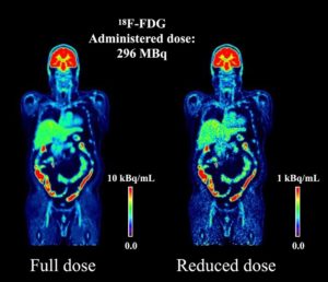
This challenge aims to develop computational algorithms capable of recovering high-quality imaging from low statistics corresponding to low dose scans, with the hope of reducing the radiation exposure to be equivalent to transatlantic flight.
Organizers:
Kuangyu Shi, Rui Guo, Song Xue, Axel Rominger, Biao Li
Respected Advisory Board Members:
Prof. Thomas Beyer, Vienna; Prof. Ronald Boellaard, Amsterdam; Prof. Simon Cherry, UC Davis; Dr. Maurizio Conti, Siemens Healthineers; Prof. Terry Jones, UC Davis; Dr. Hongdi Li, United Imaging; Prof. Nassir Navab, TU Munich; Prof. Jinyi Qi, UC Davis; Prof. Julia Schnabel, Helmholtz Center; Prof. Suleman Surti, UPenn; Prof. Kris Thielemans, UCL; Prof. Stefaan Vandenberghe, Ghent; Prof. Dimitris Visvikis, INSERM; Prof. Lifeng Yu, Mayo Clinic