
The 2020 NSS-MIC Short Course program offers 5 full-day courses and one 2 half-day course on a diverse range of topics in nuclear science and medical imaging. All courses are run by experts in their respective fields and address both theoretical foundations and practical applications. The program offered this year covers popular topics from previous years in addition to new areas, such as Radiation Hardened Design for Nuclear Measurements and Neuromorphic Computing. We encourage you to make the most of the Short Course program to diversify your background knowledge and research interests, update your skills, or simply refresh your memory.
The MIC Short Course schedule includes courses that allow participants to build their knowledge base across a variety of topics – for example, PET Kinetic Modeling and Parametric Imaging provides an excellent introduction to the kinetic modeling field for students and newcomers. Hybrid nuclear medicine devices: instrumentation and application covers physical aspects of imaging devices and their hybrid use in clinical applications. Artificial Intelligence in Medical Imaging, will provide an introduction to machine and deep learning and how it can be applied in the context of medical imaging.
This year the Short Course program runs from Saturday 31st October – Monday 2nd November, thus enabling all attendees to take part in the NSS and MIC Joint Sessions on Tuesday 3rd Nov.
For more information, please contact:
Angela Di Fulvio
Short-Course Co-Chair (NSS-Topics)
Kira Grogg
Short-Course Co-Chair (MIC-Topics)
Quanzheng Li
Short-Course Co-Chair (MIC-Topics)
SC1: Machine Learning and Neuromorphic Computing in Nuclear Science
SC 1 : “Machine Learning and Neuromorphic Computing in Nuclear Science”
Date/time/venue: Saturday 31. October 2020 at 9 AM-12.30 PM, 1.30 – 4.30 PM ET – Marina Ballroom 1
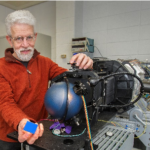
Simon Labov (Lawrence Livermore National Lab), Artur Dubrawski (Carnegie Mellon University), Jean-Roch Vilmant (California Institute of Technology) and Catherine Schuman (Oak Ridge National Lab)
Statistical methods, machine learning and artificial intelligence are quickly becoming an important part of the analysis of large data sets acquired by radiation detectors through digital electronics. These computational methods enable us to unmask data correlations, trends, and patterns that provide valuable insights on the physics phenomena under study. The methods and insights can then be used to analyze measurements with greatly enhanced performance. In this course, attendees will learn about issues with and solutions for applying machine learning to radiation measurements and then explore the application of machine learning to radiation detection. In the second part of the course, instructors will introduce functions and architectures enabled by novel neuromorphic computing devices with applications to radiation detection in nuclear physics.
Sim on E Labov, Ph. D.
Group Leader, Nuclear Security Physics
Program Leader, Nuclear Detection Program
Nuclear and Chemical Sciences Division, Lawrence Livermore National Lab
Simon Labov joined LLNL in 1987 and helped initiate a program to develop high-resolution, energy-dispersive x-ray detectors that operate at very low temperatures. Labov has held several leadership positions, including Program Leader for Nuclear Detection and Countermeasures in Global Security’s Program, Group Leader for Nuclear Security Physics in the Physical and Life Sciences Directorate, and Director of the LLNL Radiation Detection Center. Prior to joining LLNL, Labov worked as a research assistant developing instrumentation for soft-x-ray astrophysics and received a B.S. at Stanford University and a Ph.D. at the University of California, Berkeley.
Labov is an expert in nuclear detection systems, advanced spectral and multisource analysis algorithms, and distributed detector systems. He currently leads an algorithm development team for the Department of Homeland Security Countering Weapons of Mass Destruction Office ubiquitous detection SIGMA program. In 2006, he initiated research using advanced signal processing algorithms coupled to machine-learning analysis to combine heterogeneous measurements and contextual information for radiation screening operations. These techniques enable the Enhanced Radiological Nuclear Inspection and Evaluation System, now operating by the Customs and Border Protection at some of the busiest U.S. seaports, to increase sensitivity and reduce false alarms for radiation portal monitors. In 2001, he initiated the Cellular-Telephone-Based Radiation Sensor and Wide-Area Detection Network Project, which was awarded three patents and has been aggressively supported by multiple funding agencies.
 Catherine D. Schuman, Ph.D.
Research Scientist
Computational Data Analytics Group
Computer Science and Mathematics Division, Oak Ridge National Laboratory
Catherine (Katie) Schuman is a research scientist at Oak Ridge National Laboratory (ORNL). She received her Ph.D. in Computer Science from the University of Tennessee, where she completed her dissertation on the use of evolutionary algorithms to train spiking neural networks for neuromorphic systems. She is continuing her study of models and algorithms for neuromorphic computing at ORNL. Katie has a joint faculty appointment with the Department of Electrical Engineering and Computer Science at the University of Tennessee, where she co-leads the TENNLab neuromorphic research group. Katie has over 45 publications as well as six patents in the field of neuromorphic computing. Katie received the U.S. Department of Energy Early Career Award in 2019.
 Artur Dubrawski, Ph. D.
Research Professor
Carnegie Mellon University Robotics Institute
Artur Dubrawski is a Research Professor at Carnegie Mellon University Robotics Institute where he directs the Auton Lab, a group of over 80 members focusing on fundamental and applied studies into machine learning. He investigates intelligent systems that work, are useful and impactful, and studies the ways to design, build and deploy them in practice. For three decades his work has been driven by real-world applications of artificial intelligence in a range of areas including public health, food safety, health informatics, clinical medicine, intelligence, law enforcement, health of equipment, and nuclear safety. Dr. Dubrawski combines academic and real-world experience having served in executive and research lead roles in the high-tech industry.
 Jean-Roch Vlimant, Ph. D.
Research scientist
Physics Mathematic and Astronomy Department, California Institute of Technology
Jean-Roch Vlimant is a research staff member in the Physics Mathematic and Astronomy department at the California Institute of Technology. Dr. Vlimant holds a Master in Quantum Mechanics from Ecole Normal Superieure of Paris and a Ph. D. in particle physics from the Pierre et Marie Curie University. Dr. Vlimant is taking leading roles in the High Energy Physics community effort of developing deep learning and quantum computing applications for particle physics. In addition, he is actively involved with activities at the CERN, the Compact Muon Solenoid collaboration, and high-performance computing facilities worldwide.
SC2: Fast timing detectors: materials and readout
SC 2 : “Fast timing detectors: materials and readout”
Date/time/venue: Saturday 31. October 2020 at 8.30 AM- 12.30 PM ET – Marina Ballroom 2
Etiennette Auffray, Jeff Sykora, and Angela Di Fulvio
Improved detection time resolution is crucial to increase measurement precision and is enabled by new materials and fast-readout systems. This course will focus on fast timing scintillators and their applications. The first section provides an overview of well-established and novel scintillating materials for HEP, PET, materials science, nuclear energy and medical physics. The second section delves into the methods and systems for light readout, including different silicon photomultipliers technologies.
Outline
- Fast-timing scintillators and applications in HEP and diagnostic imaging (Auffray) 8.30 – 10.30 AM
- Fast-timing scintillators in neutron spectroscopy applications (Sykora) 10.30 – 11.30 AM
- Photosensors in fast timing scintillation detectors (Di Fulvio) 11.30 AM – 12.30 PM
Etiennette Auffray, Ph. D.
Senior physicist, CERN
Etiennette Auffray is a Member of the IEEE, and she has been a member of the RISC committee. She is a senior physicist at CERN, Geneva, Switzerland. She has spent over twenty-five years in the field of scintillators and their applications in particular in high energy physics and medical applications. She was actively involved in the construction of the electromagnetic calorimeter of the CMS experiment at CERN made of 75848 crystals of PWO and now in its operation. She is involved in research activities on scintillating materials for the development of PET and TOFPET through the Crystal Clear collaboration of which she is the spokesperson since 2010. In recent years she has coordinated several European projects related to scintillating materials and their applications in particular in recent years for fast timing detectors.
Jeff Sykora, Ph. D.
Senior physicist, Science and Technology Facilities Council
Jeff Sykora is a senior applied physicist at the ISIS pulsed neutron and muon source as part of the Science and Technology Facilities Council (STFC), at Harwell Oxford, United Kingdom. Jeff has 15 years of experience in luminescent materials and their applications in medical physics, charged particle spectroscopy and neutron detection. His primary focus is on neutron detectors for materials science applications. He developed neutron sensitive scintillator technology that is now in use on the single crystal diffractometer SXD, powder diffractometer IMAT and the INTER and OffSpec reflectometers at ISIS. Jeff also leads the STFC team contributing scintillation detector research to the International Collaboration for the Development of Neutron Detectors and the recently completed European collaboration, SINE2020. Lately, his research interests include new fast rise and decay time organic scintillators and nanoparticle inorganic scintillators for high count rate applications.
Angela Di Fulvio, Ph. D.
Assistant Professor, University of Illinois at Urbana-Champaign
Angela Di Fulvio is an assistant professor in the Department of Nuclear, Plasma, and Radiological Engineering (NPRE) at the University of Illinois, director of the Neutron Measurement Laboratory, and a researcher in the technical aspects of nuclear safeguards and nonproliferation. Before joining NPRE, Angela was a research scientist at the University of Michigan where she worked on radiation detection within the framework or the Consortium for Verification Technology. Her current interests include the development of detection systems for safeguards and nonproliferation applications, and techniques and algorithms for the radiation protection of the patient in radiation therapy.”
SC3: Scientific writing for journal papers and grant proposals
SC 3 : “Scientific writing for journal papers and grant proposals”
Date/time/venue: Sunday 01. November 2020 at 1:00 -3:00 PM ET – Marina Ballroom 1
Angelika Hofmann (Yale University, US)
Without clear communication, even the best data in science means little. Acquire fundamental skills of professional scientific writing and learn what constitutes clear communication in science. This workshop will navigate the complex field of professional scientific writing, covering key principles of good technical style and composition. Hands-on exercises will be included, and the important sections of manuscript and grant writing will be discussed. Emphasis will be on how to achieve maximum impact and success with the reader and reviewer in mind.
 Angelika Hofmann, Ph.D.
Strategic Projects and Communications Advisor
Office of the Vice Provost for Research, Yale University
Founder and President, SciWri Services
As Strategic Projects and Communications Advisor, Angie Hofmann works closely with Yale’s Vice Provost of Research (VPR) in coordinating the implementation of academic priorities in the sciences across the University. In her role, she also leads strategic communications efforts for the research team, is charged with advancing proposal development, and manages strategic committees for entrepreneurship and innovation.
Before joining the VPR team in 2018, Angie was Director, Corporate and Foundation Relations, Science and International Initiatives, in Yale’s Office of Development where she managed some of Yale’s most important prospects and played a key role in fundraising from leading international corporations and foundations. As part of this position, she also oversaw a team of professional scientific writers and the operational management of the Corporate and Foundation Relations office.
Angie holds a PhD in molecular biology and biochemistry from the University of California, Irvine, and was a post-doctoral fellow at the Max-Planck-Institute of Molecular Genetics in Berlin, Germany. She is renowned in the scientific writing field. She is the founder and President of SciWri Services, a venture providing editing, training, and consulting services worldwide. She is also the author of two books that have become standards in the field of scientific communication: Scientific Writing and Communication – Papers, Proposals, and Presentation (Oxford University Press, 3.ed., 2016) and Writing in the Biological Sciences (Oxford University Press 3.ed., 2018).
SC4: Integrated Circuits and Radiation Hardened Design for Nuclear Measurements
SC 4 : “Integrated Circuits and Radiation Hardened Design for Nuclear Measurements”
Date/time/venue: Sunday 01. November 2020 at 9:00 AM-12:30 PM, 3.30 – 5:30 PM ET – Marina Ballroom 2
Paul O’Connor (Brookhaven National Laboratory, US), Raffaele Giordano (University of Naples Federico II, Naples, Italy)
This one-day course is intended to introduce physicists and detector specialists to the fundamentals of integrated circuits (IC), front end design, and radiation-hardened design. The course provides an overview of analog design methodologies and semiconductor devices and then delves into the details of implementing practical circuits in modern CMOS technology. In the second part of the course, the participants will learn state-of-the-art design techniques to implement radiation hardened digital design strategies in the radiation detection readout. A basic knowledge of detectors and electronics is assumed.
9.00 AM – 12:30 PM
- Brief introduction to design methodology (CAD tools and foundry access for research-scale projects) (Paul O’Connor)
- Analog circuit design (Paul O’Connor)
- Basic architectures with elementary amplifiers (Paul O’Connor)
- Building blocks for the analog channel: charge-sensitive and pulse-shaping amplifiers, baseline stabilizers, peak detectors, track/hold, multiplexers, output stages (Paul O’Connor)
- Feature extraction: event occurrence, position, time, energy (Paul O’Connor)
- Overview of analog-to-digital and time-to-digital converters (Paul O’Connor)
- Application examples from particle physics, astrophysics, photon science, and medical imaging (Paul O’Connor)
3.30 PM – 5 PM
- Brief introduction to radiation effects in microelectronics (Raffaele Giordano)
- Radiation hardening by design (Raffaele Giordano)
- Techniques for real-time self-correction in reconfigurable ICs (Raffaele Giordano)
 Paul O’Connor, Ph. D.
Senior scientist and associate division head for the Instrumentation Division at Brookhaven National Laboratory, Upton, NY, United States
O’Connor is a senior scientist and associate division head for the Instrumentation Division at Brookhaven Lab. He attended Brown University, earning a master’s degree in electrical engineering in 1977 and a Ph.D. in physics in 1980. He joined AT&T Bell Laboratories as a member of the technical staff in 1980 and arrived at Brookhaven Lab’s Instrumentation Division in 1990. He is an author on more than 85 publications and has seven patents for microelectronic and detector technologies.
 Raffaele Giordano, Ph. D.
Assistant Professor, University of Naples Federico II, Naples, Italy
Giordano is an assistant professor in the Department of Physics at the University of Naples Federico II (Unina), Italy. He received the master’s and Ph.D. degrees in physics from the same institution in 2007 and 2010, respectively. As a postdoctoral researcher, he was involved in experiments at CERN and at the Japan High Energy Accelerator Research Organization, KEK. Since 2016, he has been an assistant professor at Unina, where he leads an R&D project on novel instrumentation for High-Energy Physics. He is an author of more than 390 scientific papers and holds two international patents for digital oscillators and radiation hardening techniques.
SC5: PET Kinetic Modeling and Parametric Imaging
SC 5 : “PET Kinetic Modeling and Parametric Imaging”
Date/time/venue: Monday 02. November 2020 at 9 AM-12.30 PM, 1.30 – 4.30 PM ET – Marina Ballroom 1
Dynamic PET imaging with tracer kinetic modeling can provide images of physiologically important parameters that have the advantages of creating higher lesion contrast, being quantitative, and allowing single tracer multiparametric imaging as compared to standard static images. While conventionally dynamic PET parametric imaging was hampered by limited scanner sensitivity and axial field-of-view, state-of-the-art commercial PET scanners now have achieved unprecedented sensitivity and also enabled simultaneous dynamic imaging of the entire body. It is becoming increasingly feasible to exploit kinetic modeling and parametric imaging for various clinical applications. This course will provide an overview of the basics of PET tracer kinetic modeling and parametric imaging. It will also cover recent advances in total-body kinetic modeling and parametric imaging. The intended audience is anyone who would like to gain a better understanding of PET kinetic modeling and parametric imaging.
Course Outline:
- Basics of compartmental modeling
- Image-derived input functions
- Reference-tissue modeling methods
- Graphical methods and linearized models
- Direct estimation of kinetic parameters
- Total-body PET kinetic modeling and parametric imaging
- Established and emerging clinical applications
Instructors:
Roger Gunn (Imperial College London, UK),
Guobao Wang (UC Davis, USA)
Marc Normandin (Massachusetts General Hospital, USA),
 Guobao Wang is an Associate Professor in the Department of Radiology, University of California Davis Medical Center. His research interest is in the advancement of PET molecular imaging using computational methods and clinical translation. The research in his laboratory commonly integrates high-resolution dynamic data acquisition with the design of advanced computational imaging algorithms (e.g., tracer kinetic modeling and image reconstruction) to derive quantitative imaging biomarkers. In close collaboration with clinicians, his research lab translates quantitative PET imaging techniques to improve clinical diagnosis, prognosis and therapy response assessment in various diseases, including fatty liver disease, heart disease, and cancer.
 Roger Gunn is Professor of Molecular Neuroimaging in the Division of Brain Sciences at Imperial College London. Professor Gunn has a background in applied mathematics and his research focuses on the application of positron emission tomography (PET) and magnetic resonance imaging (MRI) technologies to the study of pathophysiology and drug development in humans. A particular focus is the development of molecular PET probes to image targets in the brain – this includes the discovery and validation of the novel probes themselves, the associated development of quantitative analysis methods and the application of these tools in clinical studies exploring disease processes and their treatment. Professor Gunn received his undergraduate training in applied mathematics and a PhD in bio-mathematical modelling of Positron Emission Tomography (PET) data from the University of Warwick. He has worked previously at the MRC Cyclotron Unit (London, UK), held a faculty position at McGill University where he worked at the Montreal Neurological Institute before joining GSK in 2003 to help establish their Clinical Imaging Centre.In October 2011 he took up the position of Chief Scientific Officer and Head of Analysis at Imanova which is a joint venture between Imperial, UCL, Kings and the MRC targeted at providing high quality R&D imaging capabilities to the London Universities and Commercial organisations. He also holds a Visiting Professorship at Oxford University (Department of Engineering Science) and has published 120 peer reviewed journal articles in the field of imaging. Professor Gunn has served as a panel member for the MRC and on organising and programme committees at numerous conferences – he was recently Co-Chair of BrainPET 2011.
 Marc Normandin is an Assistant Professor of Radiology at Harvard Medical School. Dr Normandin’s work spans a variety of laboratory and medical imaging techniques toward the development and application of noninvasive physiological measurement technologies and assessment of therapeutic interventions. To that end, he utilizes pharmacokinetic analysis techniques, radiosynthetic/analytic procedures, and molecular biology assays to characterize biological processes in cell culture, tissue samples, and in vivo imaging in animals and human subjects. He is a recognized worldwide as a leader in molecular imaging, especially quantitative methodology for PET and MRI, and serves the scientific community through teaching locally at Harvard and MIT and internationally in the acclaimed PET Pharmacokinetics Course held annually as a satellite to the NeuroReceptor Mapping and BrainPET conferences.
SC6: Hybrid nuclear medicine devices: instrumentation and application
SC 6 : “Hybrid nuclear medicine devices: instrumentation and application”
Date/time/venue: Monday 02. November 2020 at 9 AM-12.30 PM, 1.30 – 4.30 PM ET – Marina Ballroom 2
This one-day course covers both physical aspects of SPECT/PET-CT and MR (instrumentation, requirements for integration) as well as clinical applications of hybrid nuclear medicine imaging. Basics of PET, SPECT and MR physics and instrumentation as they pertain to SPECT/CT, PET/CT, PET/MR are discussed in detail. An overview of wide range of detector technologies from Anger camera to state-of-the-art PET/MR systems will be provided.
Challenges in terms of attenuation correction, geometry integration, etc. are discussed and opportunities afforded by detection of PET/SPECT and CT or MR signals (e.g., motion compensation, partial volume correction) are explored.
We will also introduce the state-of-art deep learning method for image reconstruction and quantitation for hybrid imaging system.
These discussions will culminate with detailed assessment of what clinical applications can benefit from SPECT/CT, PET/CT or PET/MR and how to leverage its use both in the animal and human research as well as in the clinical setting.
Course Outline:
- SPECT&PET Basic Instrumentation & Image Formation (Georges El Fakhri)
- PET/MR & SPECT/PET/CT Instrumentation (Hamid Sabet)
- MR basics (Chao Ma)
- Image Reconstruction & Quantitation (Joyita Dutta)
- Clinical Applications (Georges El Fakhri)
Instructors:
Georges El Fakhri
Chao Ma
Hamid Sabet
Joyita Dutta
 Georges El Fakhri, PhD, DABR, is the Alpert Professor of Radiology at Harvard Medical School (HMS) and the founding Director of the Endowed Gordon Center for Medical Imaging at Massachusetts General Hospital and HMS with over 140 members. He is also co-Director of the Division of Nuclear Medicine and Molecular Imaging. Dr El Fakhri is an internationally recognized expert in quantitative molecular imaging (SPECT, PET-CT, and PET-MR) especially as it pertains to quantitative imaging, pharmacokinetic modeling and probing pathophysiology in brain, cardiac and oncologic PET/CT/MR. He has authored or co-authored over 200 papers and mentored over 90 students, post-docs and faculty. He has been a chartered member of many NIH study sections pertaining to Medical Imaging and Radiotherapy as well as DOD, DOE and other Foundations. He has received many awards and honors, including the Mark Tetalman Award and the E.J. Hoffman Award from the Society of Nuclear Medicine and Molecular Imaging and the Dana Foundation Brain and Immuno-Imaging Award. He was elected Fellow to the SNMMI, AAPM and IEEE for “contributions to biological imaging”.
 Chao Ma is an Instructor of Radiology at the Massachusetts General Hospital (MGH), Harvard Medical School. He received his B.S. and M.S. degree in Electrical Engineering in 2004 and 2007 both from Tsinghua University, Beijing, China. He received his Ph.D. degree in Electrical and Computer Engineering from the University of Illinois at Urbana-Champaign (UIUC) in 2013. He was the Beckman Postdoctoral Fellow of the Beckman Institute at UIUC from 2013 to 2015. In 2015, he joined the Gordon Center for Medical Imaging at MGH. Dr. Ma is a Junior Fellow of the International Society of Magnetic Resonance in Medicine (ISMRM). His primary research interests are MR spectroscopy/spectroscopic imaging, cardiac MRI, MR RF pulse design and quantitative PET/MR.
 Hamid Sabet is an Assistant Professor of Radiology at Harvard Medical School, and faculty at Gordon Center for Medical Imaging and Nuclear Medicine and Molecular Imaging Division at Massachusetts General Hospital. He earned his PhD in Quantum Science and Energy Engineering from Tohoku University, Japan in 2008. After a postdoctoral training at Rush Medical Center in Chicago (2009-2010), he worked as staff scientist at RMD Inc. from 2011 to 2014. He joined MGH/Harvard as faculty in 2014 and established Radiation Physics and Instrumentation Lab in GCMI, Radiology department. The research theme of his Lab is development of novel detector technologies for nuclear medicine and x-ray imaging, and image-guided surgical applications.
 Joyita Dutta is an Associate Professor in the Department of Electrical and Computer Engineering at the University of Massachusetts Lowell, where she directs the Biomedical Imaging and Data Science Laboratory (BIDSLab). She also holds faculty appointments at Harvard Medical School and Massachusetts General Hospital. Dr. Dutta received her B.Tech. (Honors) in Electronics and Electrical Communication Engineering from the Indian Institute of Technology Kharagpur, India, in 2004 and her M.S. and Ph.D. degrees in Electrical Engineering from the University of Southern California in 2006 and 2011 respectively. Her research interests are machine learning and signal processing for image, graph, and time-series datasets with an emphasis on multimodal information integration. In 2013, she received a Young Investigator Award from the SNMMI Computer and Instrumentation Council. She was also a recipient of the SNMMI Mitzi & William Blahd MD Pilot Research Grant (2013-2014), the American Lung Association Senior Research Training Fellowship (2013-2015), and an NIH K01 Career Development Award (2015-2020). Her contributions to medical imaging have been recognized by the 2016 Tracy Lynn Faber Memorial Award from the SNMMI and the 2016 Bruce Hasegawa Young Investigator Medical Imaging Science Award from the IEEE.
SC7: Artificial Intelligence in Medical Imaging
SC 7 : “Artificial Intelligence in Medical Imaging”
Date/time/venue: Tuesday 03. November 2020 at 9 AM-12.30 PM, 1.30 – 4.30 PM ET – Marina Ballroom 1
This is an introductory artificial intelligence course with a strong focus on medical imaging. The objective of the course is to provide an entry level knowledge of machine learning and deep learning, how it fits into medical imaging, in what situations it is the appropriate tool for clinical applications.
Course Outline:
- Introduction to deep learning
- Hardware and software for deep learning
- Deep learning for medical image reconstruction
- Deep learning for detection and classification in medical applications
- Deep learning for image segmentation
Instructors:
Quanzheng Li
Kuang Gong
Kyungsang Kim
 Quanzheng Li is an Associate Professor of Radiology at Massachusetts General Hospital, Harvard Medical School. Dr. Li is also the scientific director of MGH/BWH Center for Clinical Data Science, the director of the Center for Advanced Medical Computing and Analysis, and a Core faculty of Gordon Center for Medical Imaging, Massachusetts General Hospital, Harvard Medical School. He received his B.S degree from Zhejiang University in 1997, M.S. degree from Tsinghua University in 2000, and his Ph.D degree in Electrical Engineering from the University of Southern California (USC) in 2005. He did his post-doctoral training at USC from 2006 to 2007, and was a Research Assistant Professor from 2008 to 2010. In 2011, he joined the Radiology Department at Massachusetts General Hospital. Dr. Li is the recipient of 2015 IEEE Nuclear and Plasma Sciences Society (NPSS) early achievement award. He is an associate editor of IEEE Transaction on Image Processing, IEEE Transaction on Medical Imaging, and members of editorial boards of Theronostics and Physics in Medicine and Biology. Dr. Li has more than 140 peer reviewed articles and his team has won AAPM-NIH low dose CT challenge and 2018 Camelyon Challenge on digital pathology. His research interests include image reconstruction and analysis in PET, SPECT, CT and MRI, and artificial intelligence in health and medicine.
 Kuang Gong is an Instructor of Radiology at Massachusetts General Hospital and Harvard Medical School. He received his B.S degree in Information Science and Electronic Engineering from Zhejiang University in 2011, M.S. degree in Statistics and Ph.D. degree in Biomedical Engineering from UC Davis, in 2015 and 2018, respectively. His research interests include medical image reconstruction, PET physics, as well as machine learning applications in PET imaging. He currently focuses on deep learning-based PET image reconstruction, attenuation correction and parametric imaging.
 Kyungsang Kim is an instructor of Radiology at Massachusetts General Hospital (MGH), Harvard Medical School (HMS). He received a Ph.D. degree in Dept. of Bio and Brain Engineering at Korea Advanced Institute of Science and Technology (KAIST) in 2014. He was a postdoctoral research fellow at the Massachusetts General Hospital (MGH) and Harvard Medical School (HMS) from 2014 to 2017. His research topics are image reconstruction and analysis using compressed sensing and deep learning for medical applications such as low dose CT reconstruction, PET/MR joint reconstruction, and PET parametric imaging.